At MIT, researchers have cracked a problem that has frustrated ultrasound technicians for decades: the cognitive leap from reading flat, two-dimensional images on a screen to mentally reconstructing them into a three-dimensional picture of what's actually inside a patient's body. Now, wearing a virtual-reality headset, they can see precisely what lies beneath the skin in real time — a breakthrough that could transform how medical ultrasound is learned, taught, and performed in clinics around the world.
The challenge is real. Ultrasound works by bouncing high-frequency sound waves off tissue, capturing the echoes as 2D slices that technicians must painstakingly reassemble in their minds. It's a skill with a steep learning curve, and the mental strain — what researchers call "the mental tomography bottleneck" — can lead to missed details and diagnostic errors. MIT graduate students Jason Hou and Shrihari Viswanath, working under the supervision of Canan Dagdeviren, an associate professor of media arts and sciences at MIT, set out to remove that cognitive burden entirely.
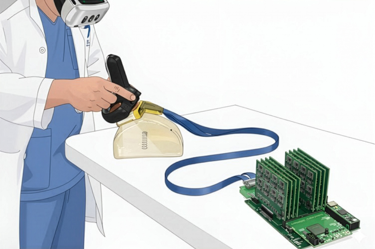
Their solution combines two existing technologies in a novel way: real-time 3D ultrasound imaging and augmented reality. The team developed AR-VIU (augmented real-time volumetric imaging in ultrasound), a system built around an ultrasound probe slightly smaller than a deck of cards. The probe uses a chirped data acquisition system and contains an array arranged in the shape of an empty square — a design that captures 3D images with fewer ultrasound elements than traditional systems, making it less expensive and power-hungry to build.
The probe streams its data to Unreal Engine, a 3D graphics platform, which converts the raw ultrasound information into a precise three-dimensional rendering without losing any detail. When a user puts on an AR/VR headset, they see this 3D digital ghost image superimposed over the actual object being scanned, as if they had X-ray vision. By tilting their head or moving around, they can view the internal structure from any angle they choose.
To test whether this actually worked, the researchers studied 18 participants — nine experienced ultrasound experts and nine complete novices. Each performed identification tasks with four different imaging methods: standard 2D images on a screen, 3D images on a screen, AR-VIU in 2D, and AR-VIU in 3D. In one test, they tried to identify hidden objects (springs, balls, screws) inside opaque gelatin containers. In another, they used a pen to mark the exact spot where a biopsy needle should go, simulating a critical clinical task. The results were striking: AR-VIU significantly improved everyone's ability to identify and locate objects, with the biggest gains among inexperienced users, whose performance approached that of experts.
Dagdeviren believes the implications extend far beyond the research lab. "For training, this could make ultrasound more intuitive and more understandable," she says. "On the clinical side, it could be less time-consuming, more accurate, and also give health care providers more peace of mind. They wouldn't have to wonder if they missed anything." The research, published in Nature Communications Engineering, involved collaboration with Bowen Wu and two MIT Summer Research Program students, Cinay Dilibal from Dartmouth College and Tanisha Shende from Oberlin College. While 3D ultrasound has long been used in specialized fields like fetal imaging and heart scanning, it remains expensive and rare. AR-VIU's simpler, cheaper design could finally make 3D ultrasound practical for hospitals everywhere — potentially speeding up training, reducing diagnostic delays, and improving the safety of procedures that rely on precise needle placement.