Kurt Schilling stood at the threshold of a discovery that had eluded neuroscience for decades: the first complete map of how the human brain's white matter grows, changes, and ages across an entire lifetime. For the first time, researchers at Vanderbilt University have charted 72 distinct neural pathways from birth to 100 years old, using data from nearly 42,000 brains—more than 4 million individual MRI images analyzed through an advanced AI-enabled computing platform.
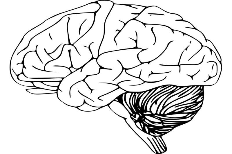
White matter, the bundled nerve fibers that transmit signals throughout the brain and body, has long been poorly understood at the population level. Unlike height and weight charts that have guided pediatricians for generations, neurology had no comparable tool to track neural development or flag abnormalities before disease takes hold. That absence meant doctors couldn't easily detect early signs of Alzheimer's, Parkinson's, epilepsy, or autism spectrum disorders—conditions where white matter dysfunction plays a central role.
The Vanderbilt team's findings fundamentally reshape our understanding of how the brain evolves. Rather than developing as a unified whole, the brain's 72 major white matter pathways follow strikingly independent timelines. Overall cerebral white matter volume expands rapidly during early development, peaking in a person's early to mid-30s before beginning a gradual decline. But pathway integrity—a key marker of white matter organization—matures far earlier, rising quickly through childhood and adolescence before plateauing in the mid-20s and then steadily declining. This means different brain circuits associated with different functions are vulnerable at different life stages, a pattern that opens entirely new avenues for understanding both typical development and disease.
Michael Kim, the lead author and a Vanderbilt Ph.D. student in computer science, emphasized the practical implications: "Defining these pathway-specific trajectories and milestones allows researchers to explore interesting neurobiological questions. They also help us investigate how white matter abnormalities present similarly or differently across diseases." That ability to detect abnormalities early—before symptoms emerge—could transform early intervention in neurodegenerative conditions and developmental disorders alike.
The research represents nearly two decades of methodical collaboration between Vanderbilt University and Vanderbilt Health. Bennett Landman, University Distinguished Professor in Electrical and Computer Engineering and Radiology, spent roughly a decade harmonizing MRI data across 50 population studies, a foundational effort that made this discovery possible. As Landman noted, "What's exciting is that these data harmonization efforts are reaching the point of enabling transformative discovery."
The implications ripple across the full spectrum of neurological disease. As Schilling observed, "There's not a single neurodegenerative disease that doesn't implicate white matter dysfunction in some way." With these growth charts now established, researchers can finally investigate how white matter abnormalities emerge in autism, ADHD, dyslexia, multiple sclerosis, and dozens of other conditions—each against a backdrop of what healthy development actually looks like across the lifespan.
Published in Nature, this work offers neurologists a new diagnostic lens and gives researchers a precise reference point for understanding how the brain changes across a human lifetime. For patients and families facing neurological disease, it means the possibility of earlier detection, more targeted interventions, and a clearer picture of what normal aging looks like.