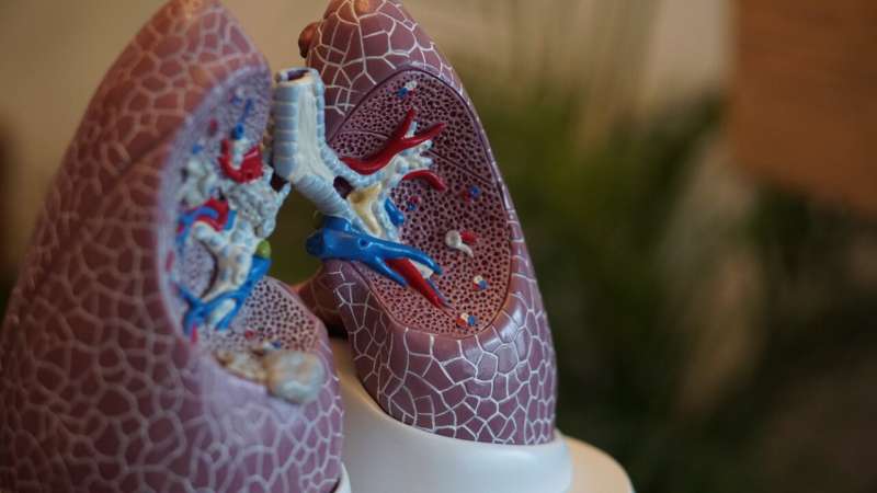
At nine major medical centers across the United States, doctors performing lung biopsies have discovered a tool that could change how they diagnose lung disease: a fingertip-sized cryoprobe that freezes tissue instead of crushing it.
The findings matter because an accurate lung biopsy can mean the difference between a swift diagnosis and weeks of uncertainty—and for patients with cancer, infections, or transplant complications, time is everything. When a bronchoscope slides through a patient's airway to collect a tissue sample, the quality of that sample determines whether doctors can confidently identify what's happening in the lungs. A damaged or incomplete specimen can lead to repeat procedures, delayed treatment, and unnecessary anxiety.
In the FROSTBITE-2 randomized trial published in JAMA, researchers led by Dr. Fabien Maldonado at the Vanderbilt Lung Institute compared two biopsy methods on 500 patients. When doctors used the newer 1.1-millimeter cryoprobe—a device that uses localized freezing to extract tissue—the diagnostic yield reached 88.6%. When they used traditional 2.0-millimeter forceps, which pinch off tissue but can crush it in the process, the success rate was 78.8%. That nearly 10-percentage-point difference is substantial in clinical terms. The gap widened significantly for patients with pulmonary nodules or masses: 83.2% diagnostic accuracy with the cryoprobe versus 70.1% with forceps.
What makes the cryoprobe particularly elegant is its size. Unlike larger cryoprobes tested in previous studies, this 1.1-millimeter version is small enough that doctors can remove the tissue sample through the working channel of the bronchoscope without withdrawing the entire scope. That design choice has a cascading benefit: it enhances safety by reducing trauma to the airway.
The safety data underscore why this matters. In the forceps group, four patients experienced pneumothorax—a collapsed lung requiring a chest tube to re-expand it. That's a 1.6% complication rate. The cryoprobe group had zero pneumothoraces. Neither group experienced significant bleeding or respiratory failure, suggesting both methods are fundamentally safe when performed by experienced interventional pulmonologists, but one clearly carries less risk.
Dr. Maldonado, who directs the Interventional Pulmonology program at Vanderbilt and serves as vice chair of the Interventional Pulmonary Outcomes Group, framed the work in patient-centered terms: "A structurally intact, sufficiently large tissue sample from a targeted area in the lung increases the likelihood of an accurate diagnosis." He noted that accurate diagnoses delivered quickly help patients access treatment faster—a principle that applies across lung cancer, transplant medicine, and interstitial lung disease.
The trial was conducted at nine medical centers specializing in high-volume transbronchial biopsy, ensuring the results reflect real-world expertise. It represents the work of an international collaborative of clinical experts dedicated to improving outcomes through rigorous, multicenter research.
This finding sits within a larger momentum: researchers are actively investigating the next generation of this technology through FROSTBITE-3, already underway. Each refinement brings patients closer to diagnosis that is simultaneously more accurate, safer, and faster—a rare alignment of better outcomes across multiple dimensions.