Every human cell wears a coat of sugar molecules so intricate that scientists are only now learning to read it like a secret language. This gossamer layer, called the glycocalyx, has always been there—wrapping every cell in your body in a protective embrace—but researchers at Germany's Max Planck Institute for the Science of Light have just developed the tools to map it in stunning detail. Their breakthrough, published in Nature Nanotechnology, suggests that the patterns these sugars make on a cell's surface could one day become an early warning system for cancer and other diseases.
The glycocalyx isn't static. These complex sugar molecules are constantly shifting and reorganizing, responding to what's happening inside the cell. Professor Leonhard Möckl and his team in the institute's "Physical Glycosciences" research group became fascinated by the question: What if this surface layer isn't just protective, but informative? What if it acts like a display screen, broadcasting a cell's internal state to the outside world?
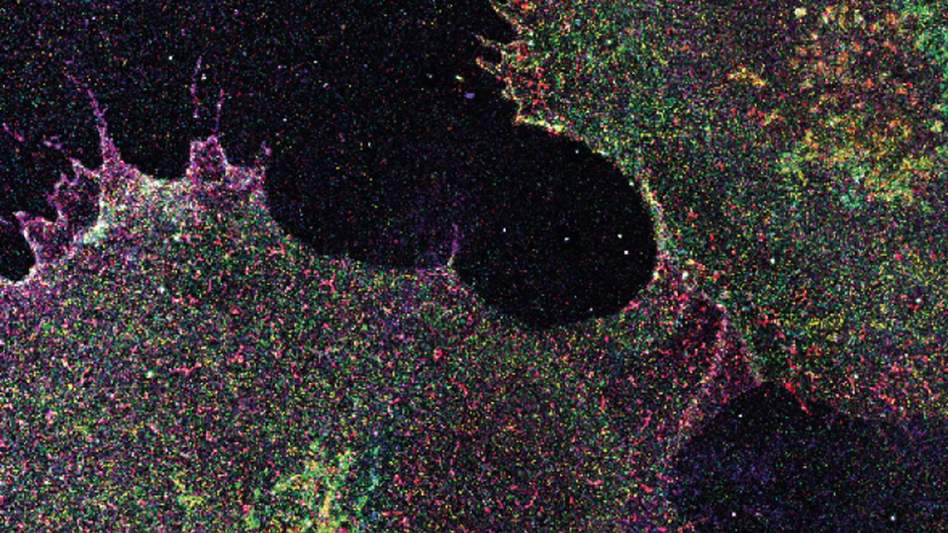
To find out, they developed a technique called "Glycan Atlasing," combining cutting-edge super-resolution microscopy with careful mapping at the level of individual sugar molecules. Working across cell culture lines, primary human blood cells, and actual tissue samples, they created the first detailed atlases of these nanoscale sugar structures. What they discovered was striking: the glycocalyx changes its molecular arrangement depending on the cell's condition. Immune cells showed visibly different sugar patterns after being stimulated, just as they would during an immune response. For the first time, researchers had direct evidence that cells were essentially displaying their internal information on their surface.
But the most promising finding may be medical. The team's measurements showed that these sugar patterns could reliably distinguish between different cellular states with a precision that surprised even experienced researchers. They could identify separate stages of cancer development. They could tell the difference between activated and inactive immune cells. And they could distinguish cancerous regions from healthy regions in samples of human breast tissue—a proof-of-concept that suggests the method might work in real clinical settings.
"The results provide a promising foundation for the development of future diagnostic methods, as Glycan Atlasing delivers reliable results even in complex samples," Möckl explains. What makes this particularly significant is that the technique works not just in pristine laboratory conditions, but in messy, complicated tissue samples closer to what doctors actually deal with in hospitals.
The team now faces the exciting work of scaling up. They plan to expand the method by analyzing additional target structures and automating more of the process. They want to study much larger numbers of samples so the technique can eventually move from the laboratory into routine medical practice. As Möckl outlines the future, there's a clear sense of purpose: "In large-scale studies, we want to investigate which surface patterns are associated with specific disease courses or therapeutic responses and how cell states can be detected early and objectively via the surface."
If they succeed, the implications could reshape early disease detection. Imagine a diagnostic test that reads the sugar language on your cells, identifying cancer or other illnesses before symptoms appear. The Max Planck team has cracked open the code. Now comes the harder work of teaching medicine to speak the same language.