Nestled in Cambridge laboratories, tiny pea-sized clusters of human brain tissue are rewriting what scientists thought they knew about spinal cord injury. Dr. András Lakatos and his team at the University of Cambridge have created miniature versions of the connected brain and spinal cord system—complete with functioning neural circuits—and their findings suggest that nerve damage long dismissed as permanent may actually be reversible.
The question has haunted neuroscience for decades: why does the adult nervous system lose its ability to repair itself? When the human brain and spinal cord suffer damage, the axons—the long nerve fibers that carry movement signals from brain to muscle—rarely regrow. This biological dead end explains why spinal cord injuries so often result in permanent paralysis, and why conditions like motor neurone disease and multiple sclerosis inflict lasting disability. Understanding when and why neurons stop regenerating could unlock treatments for conditions previously considered untreatable.
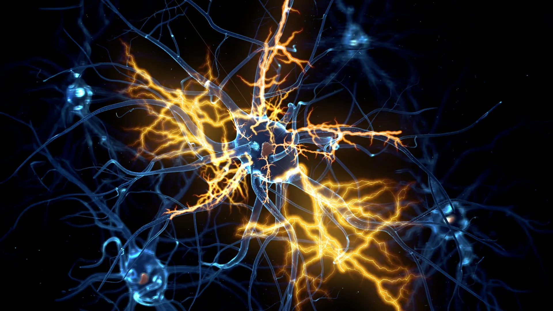
In 2021, Lakatos's team developed their first brain organoids from patient-derived stem cells. Now, in a study published in Cell Reports, they've taken the work further by building a system that mimics the actual connection between brain and spinal cord. The researchers kept the two tissue types physically separate in the lab, then watched as axons grew across the gap and linked up with spinal cord tissue. Remarkably, the resulting neural circuit was functional enough to trigger actual muscle contractions—proof that their miniature system genuinely replicated how the body's movement signals work.
What they discovered by maintaining these systems for over a year fundamentally changed the picture. Until roughly day 150 of development—around the middle stage of pregnancy—damaged axons could still regrow. After that point, neurons showed a sharp decline in their regenerative ability. "Neurons taken from less mature organoids regrew long fibers after injury, but those from more mature organoids showed a sharp drop in their ability to regrow," explained George Gibbons, first author of the study. "In other words, poor regeneration is built into human neurons as they mature in the central nervous system."
The team identified a network of genes acting like a biological switch that progressively limits axon growth as neurons mature. But here's where hope enters the picture: when researchers blocked key regulators within this network, the neurons regained their ability to grow axons again. The breakthrough deepened when the scientists screened a database of drug compounds and found that lynestrenol—a hormone medication already approved for menstrual disorders and contraceptive use—significantly improved axon regrowth in damaged neurons.
Lakatos emphasizes that lynestrenol itself may not be the final answer to spinal cord repair, but its effectiveness proves the principle: human neurons can be directly targeted to regenerate their axons. "Although we still need to show that this strategy will also help to re-establish appropriate connections between the brain and spinal cord cells," he said, "this gives us hope that one day we may be able to treat conditions previously thought untreatable."
The implications extend beyond one drug. By using human organoids rather than relying solely on animal models, these researchers have uncovered biology unique to human neurons—biology that animal studies might easily have missed. That biological specificity could be the missing link that transforms how researchers approach neurological disease.