Across 12 human brain tumors, researchers mapped more than a million cell nuclei and hundreds of thousands of tissue locations to investigate how glioblastoma cells become diverse. Their atlas, called GBM-space, brings together detailed measurements from tumors that are often studied as if their cancer cells were one uniform group.
Glioblastoma is an aggressive adult brain cancer that remains incurable. Its cells can occupy very different states: some resemble developing brain cells, while others show patterns linked to tissue injury. These differences may help explain why tumors respond unevenly to treatment. Earlier work also suggested that cancer cells can shift between states, but it was unclear whether they followed a shared route or changed in different ways from tumor to tumor. Finding a recurring route could point researchers toward a weakness shared by many tumors.
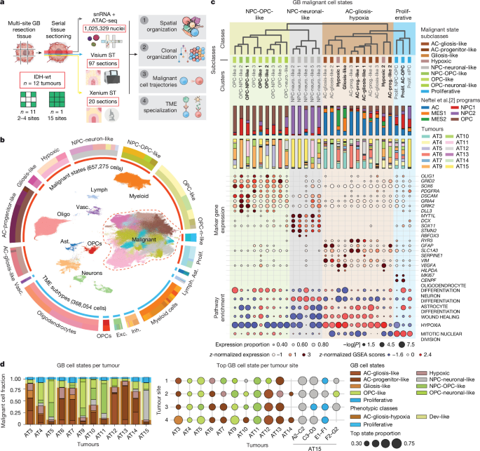
To build the atlas, the team studied 12 primary glioblastomas without mutations in the IDH gene, a group known as IDH-wild-type. They sampled 57 sites across the tumors, taking four sites in most cases and as many as 15 in one tumor. At each site, they paired single-cell measurements with maps showing where cells sat in the tissue. The methods let researchers examine both which genes were active and how accessible parts of the cells' DNA were, alongside the cells' locations.
The resulting dataset included 97 spatial tissue sections and more than 300,000 spatial transcriptomes, measurements of gene activity tied to positions in tissue. In total, the atlas contained 657,275 malignant cells and 368,054 cells from the tumor microenvironment: the surrounding immune, support and blood-vessel cells. The team also used other methods, including tissue staining and whole-genome sequencing, at selected sites. The data are available through the GBM-space website, where other researchers can explore the tumor maps.
The study reports that glioblastoma cells follow a stereotyped, or recurring, spatiotemporal trajectory as the tumors develop their varied cell states. That finding links cell identity, tumor location and change over time in one picture. The supplied study details do not specify the exact sequence of states in that trajectory, so its practical meaning for treatment remains a question for further research. Still, an atlas spanning many sites and patients gives scientists a way to test whether the same patterns appear in other tumors and to study how surrounding tissue may shape cancer cells.
For patients, the work is not a new treatment, and it does not make glioblastoma less serious. Its value is a clearer map of a difficult disease. If researchers can pin down which steps in the shared trajectory matter most, they may eventually find ways to interrupt cancer cells as they shift into states that help tumors survive.